![]()
The SAGE Spectrometer
![]()
![]()
![]()
![]()
![]()
![]()
![]()
![]()
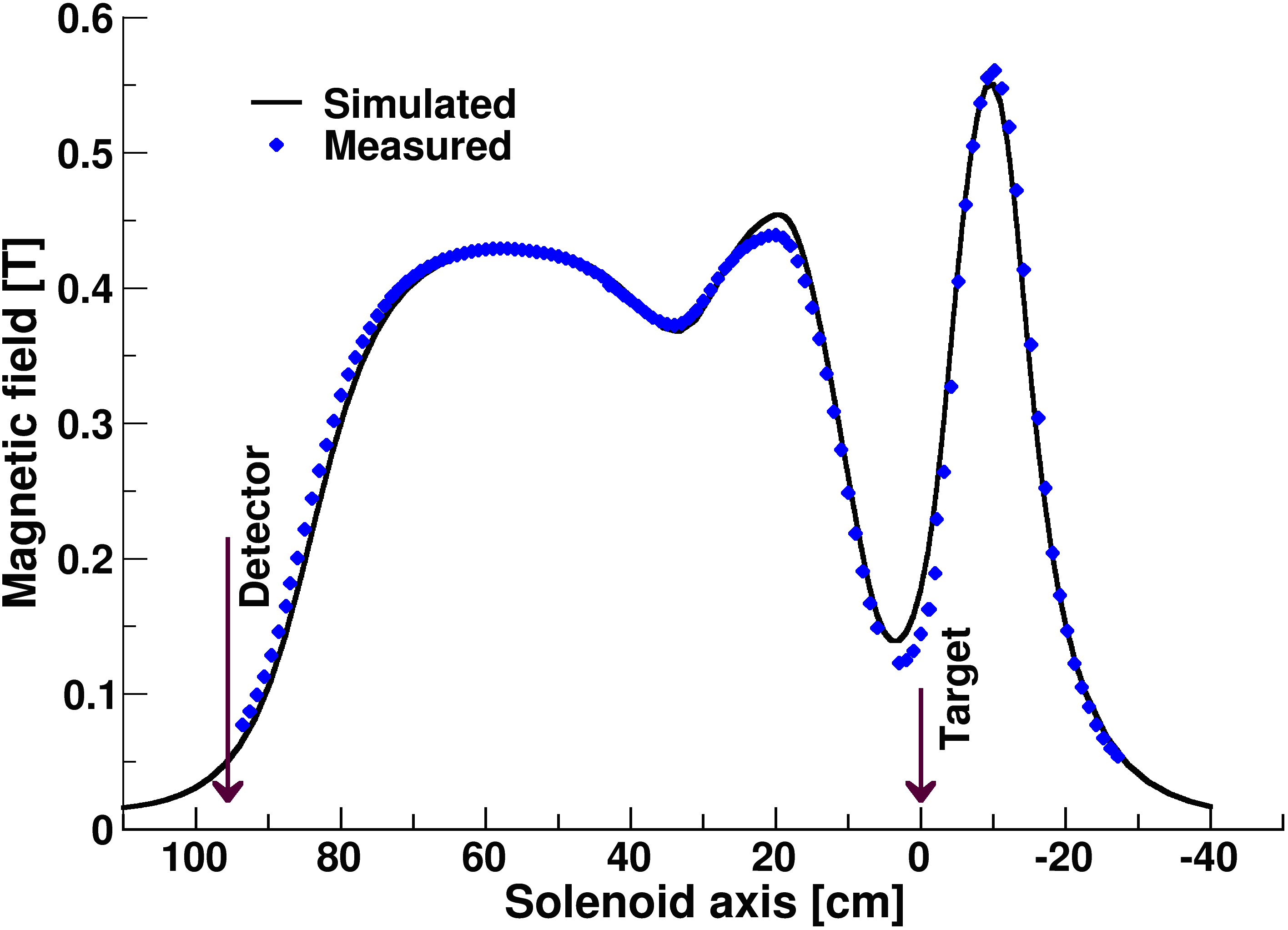
| The electrons are transported away from the hostile target region and towards the silicon detector by a magnetic field. The field is induced by three solenoid coils designed in a way which maximizes electron transmission without compromising γ-ray efficiency. |
 |
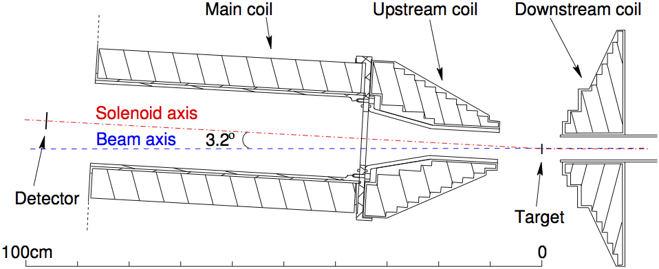 |
Nearly collinear geometry between the solenoid coil and beam axes reduces Doppler broadening. Positioning the silicon detector upstream of the target reduces the δ-electron flux which reaches the detector. |
| A high-voltage barrier positioned between the target and detector further reduces the δ-electron flux. |
 |
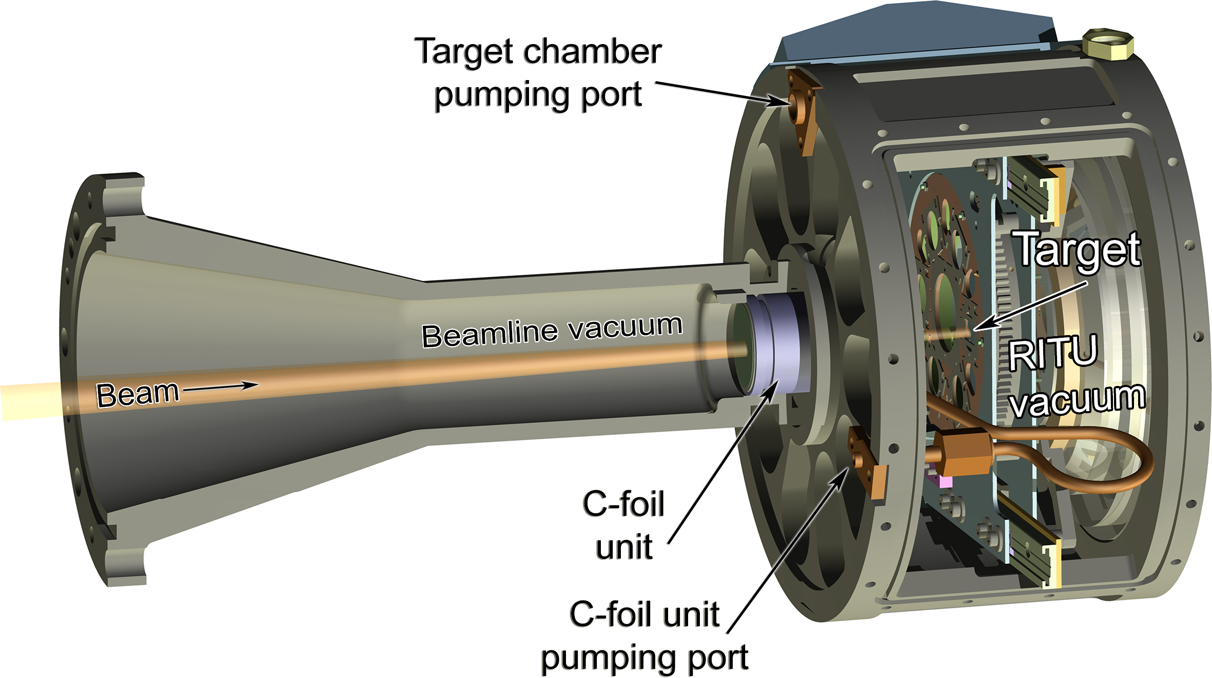 |
A dual carbon-foil system is used to separate the high-vacuum region of the high-voltage barrier from the target region. For target cooling purposes the target chamber is in the same vacuum as RITU, which operates with helium pressures up to 1mbar. |
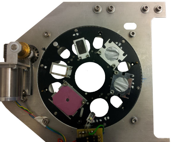
| Up to six targets can be positioned simultaneously on a target wheel. The same target wheel offers the possibility to use a larger rotating target. |
 |
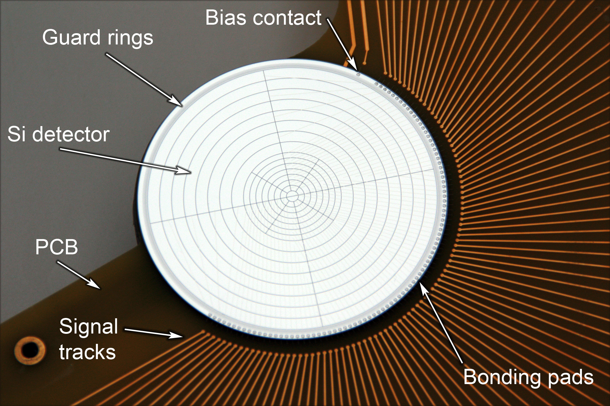 |
The electrons are detected by a 1mm thick silicon detector segmented into 90 individual pixels. |
| The detector is mounted on a PCB which also accommodates the preamplifiers. | 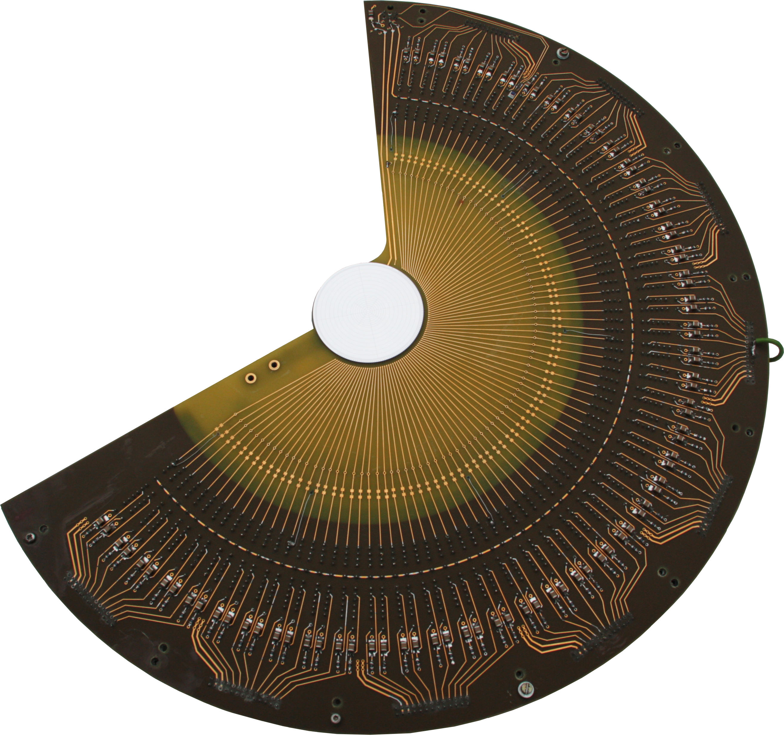 |
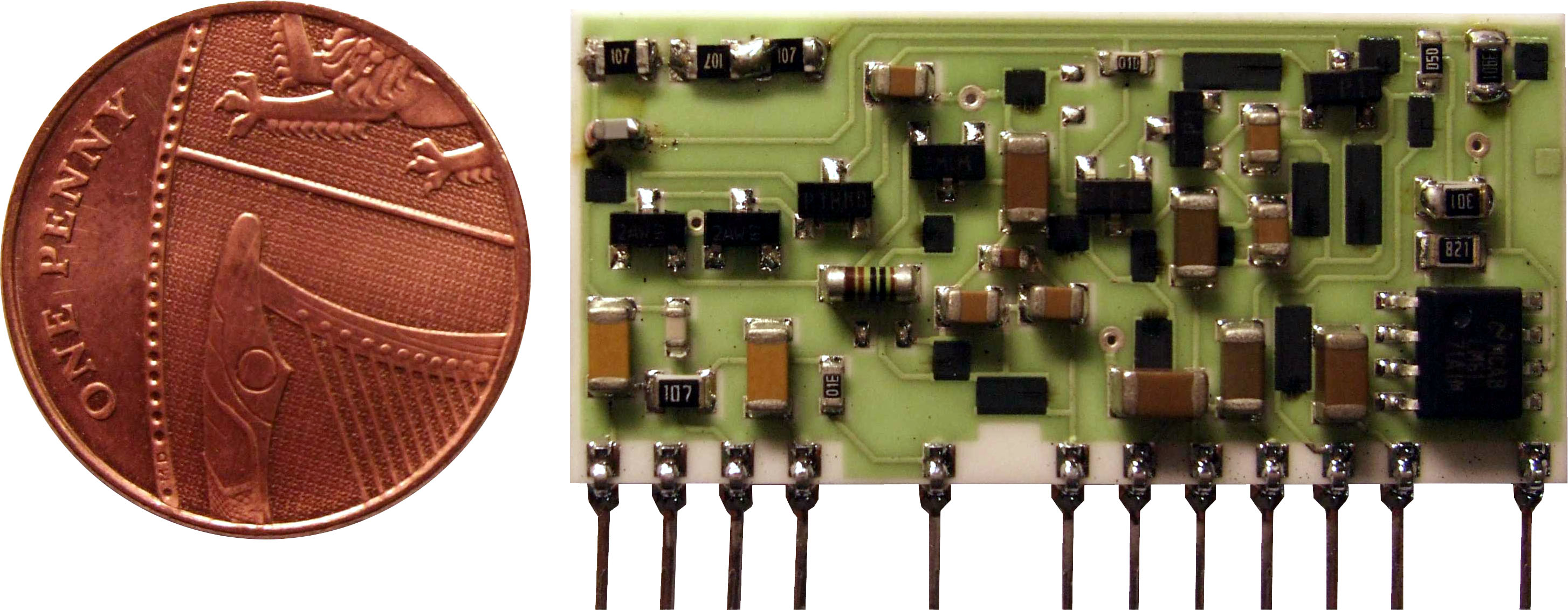 |
Each detector pixel is connected to a CAEN A1422 charge sensitive hybrid preamplifier. |
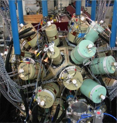
| The γ rays are detected by the JUROGAM II array which consists of 24 four-fold segmented clover detectors and 15 Eurogam Phase I-type detectors. When used with SAGE, 5 of the Phase I-type detectors are removed. All the germanium detectors are Compton-suppressed. Further information on JUROGAM II can be found here. |
 |